The dark-field microscope itself does not improve the resolution physically. What it improves is the contrast of the edges originally covered by the bright field illumination light. This is my understanding of the dark-field microscope. I haven’t studied the detailed mathematical principle of the dark-field microscope for the time being. I’m not sure whether my understanding is correct or not. Let’s use the light path diagram to explain it descriptively. If there is any error, please correct it.
The realization principle of dark-field microscope is very simple. It is to add a light mask in front of the condenser, and even write on the official website of Olympus that it can be realized with a coin. However, the effect of its implementation is impressive: it enhances the contrast of the edge part of the object, a bit like edge recognition in digital image processing. Therefore, it is very practical to image many fibrous samples that cannot be seen by ordinary open-field microscope, such as bacterial flagella, which can be seen clearly without staining.
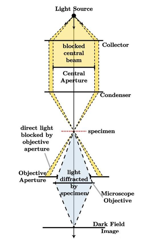
The above is the schematic diagram of the light path of the dark-field microscope in Wikipedia. The experience of light in the microscope is:
1. The point light source emits spherical light, which is shrunk to plane light through a condenser;
2. Through the barrier, the plane light is blocked in the middle, forming a very narrow ring light source;
3. The annular illumination light is then contracted by a condenser, and the sample is placed at the focal plane of the condenser to make the illumination light pass through;
4. Most of the illumination light directly passes through the focal plane without scattering with the sample, and then is blocked by the aperture in front of the objective lens;
5. A small part of the illuminating light scatters with the sample, and the direction changes. Some of the scattered light can pass through the aperture and enter the objective lens, so as to image.
This is the general imaging principle of the dark-field microscope, which is to process the illumination light into a ring and pass through the sample at a large incidence angle, so that the bright background illumination light can be isolated later, so that the originally dim details can be highlighted and the contrast can be improved. However, the imaging depends on scattered light, so the imaging is mainly at the edge and other places with obvious scattering. The uniform part of the sample is directly penetrated by the illumination light, and the imaging is not clear.
The picture below is the image of a group of researchers using dark field when studying bacterial flagella. It can be clearly seen that the dark field basically only images the edge of the sample, and the background is completely black (because the illumination light is isolated). The shape of bacterial flagella in the picture is so clear, which cannot be achieved by direct observation with a light field microscope.